ENDOSCOPIC IMAGES
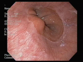
Normal GE junction
This is the gastroesophageal junction, or the location where the esophagus meets the stomach. The glistening white appearance in the foreground is the esophagus, and this is entirely normal in these patients. The esophagus meets with the stomach which is the salmon color and that also is normal. You can see that where they meet it is somewhat irregular, and it is called the Z-line. The Z-line, as you can tell from its name, is quite irregular and that is entirely normal. Often when people get heartburn, we see more irregularities of the Z-line, or we see inflammation or irritation above the Z-line in the esophagus.
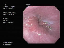
Normal gastroesophageal junction
This is simply another example of a gastroesophageal junction showing the transition from the esophagus to the stomach. This is completely normal.
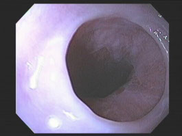
Esophageal web
This photograph is one of the bottom portion of the esophagus, which is narrowed. There is a web at this level. Often people are born with these webs, and when they are very tight, they can cause difficulty swallowing food. If that is the case, we can stretch them out with what we call dilators, which allows the food to pass more easily. This is felt to be a completely benign condition, and other than being troublesome, with difficulty swallowing food, it does not cause any serious problems.
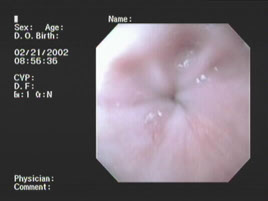
Achalasia
This is the very bottom of the esophagus, and compared to the photograph we saw in gastroesophageal junction, this is very tightly shut; in fact, there is only a pinhole opening. In patients with this condition, achalasia, there is difficulty in swallowing food and it backs up into the esophagus. We often have to operate on patients to open this area up, but there are more recent therapies one of which is shown in the next photograph.
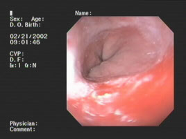
Achalasia after Botox injection
This is the same patient in whom we saw achalasia, but we have injected Botox, which is used for many different medical and cosmetic purposes, it is in the very bottom of the esophagus where the muscle was extremely tight. Botox stops the muscle from being quite as tight and allows it to open up. This effect is temporary, but typically lasts several months and gives remarkable relief of the symptoms. If you simply compare the opening in this patient, you can see a dramatic difference.
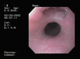
Mid-esophageal diverticulum
This is a mid esophageal diverticulum, or out-pouching. It typically causes no symptoms, it is not precancerous, and is not terribly uncommon.
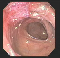
Ischemic colitis
This is a photograph taken in the colon of an area of ischemic colitis. This condition occurs when there is a lack of blood flow to the superficial cells of the colon and thus many of them die. There is often bleeding, generally there is no pain, and what we see here is an ulcer which is healing. This condition tends to get better on its own, and in this patient, there clearly is healing.
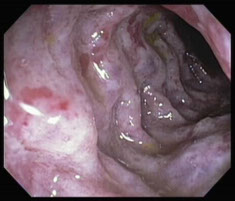
Ischemic colitis-severe
This is a photograph of another patient with ischemic colitis but as compared to the other photo, we can see severe redness, swelling, and almost a bluish appearance to the wall of the bowel and as opposed to being along only one wall, this is almost the entire bowel. This is a very severe case of ischemic colitis. It may and may not heal and often when it does heal, it heals with scarring. This is obviously a serious condition.
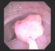
Polyp being snared
This is a large polyp in the colon. A polyp is a growth of tissue that can grow and become cancerous and, in fact, when this polyp was examined under the microscope, there were some cancerous cells in it. Polyps need to be removed, and that is why we do colonoscopies.
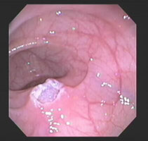
Polyp cautery site
This is the site where the previous polyp has been removed. We use a snare, which has a looped wire on the end, to grab the polyp. We then apply electrical current to burn off the polyps that there is no bleeding. From this photograph, we can see that the burn is quite deep and flushed with the wall of the colon. It was necessary to do this to get the entire polyp.
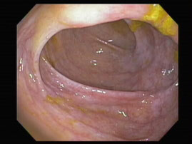
View of ileocecal valve in cecum
This is a photograph of the ileocecal valve, which is seen towards the top of the photograph, and the cecum. The ileocecal valve is where the small intestine empties into the large intestine and there is a valve behind the fold that we are looking at. You can see that this fold is thicker than the fold on the bottom of the screen where there is no such valve. In the distance, we see the end of the colon, which is otherwise known as the cecum. This is completely normal.
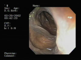
Melanosis coli
This is the photograph of the colon, and there is an extremely dark appearance to the wall of the colon. This is seen in patients who have taken laxatives over many years and the pigment from the laxative gets deposited in the wall of the bowel giving an extremely dark appearance to it. This is a benign condition, not cancerous, and does not become cancerous, but often it is quite obvious.
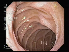
Large hiatal hernia
This is a large hiatal hernia. A hiatal hernia is a portion of the stomach that has been pulled up into the chest above the breathing muscle, or the diaphragm. Normally, the stomach is entirely in the abdomen, though when pulled above that muscle, which separates the chest from the abdomen, a bit of the stomach is in the chest. In this case, the hiatal hernia appears to be entirely normal, in that the tissue looks completely normal. This is an extremely common condition, and may make it more likely that one who has it develops heartburn.
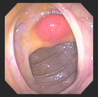
Reddened ileocecal valve
This is the ileocecal valve, or the valve where the small intestine leads into the colon. There is a very large reddened area, and underneath it we see a yellowish appearance. This is simply mildly inflamed, with a large amount of fatty tissue, which is completely benign, below it. This is not a precancerous condition.
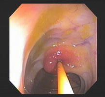
Reddened ileocecal valve after biopsy with fatty tissue extruding
In this photograph we have taken a biopsy forceps, it is closed, and I have pushed it into the valve. We can see how easily this looks like a pillow and we are pushing it in one spot. It is extremely soft, not hard, and this is very characteristic of lipomas, or areas of fatty tissue. Again, this is completely benign.
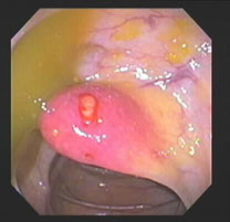
Reddened ileocecal valve after biopsy with fatty tissue extruding
This is the same valve that we have seen in the prior two photos, and a biopsy has been taken. We can see some yellowish tissue coming from where the biopsy was taken which is in the center of the valve. There is a very tiny amount of blood around it and there is fatty tissue, which is starting to extrude from the biopsy site. The biopsy was completely benign, and confirmed that this was, in fact, a large lipoma.
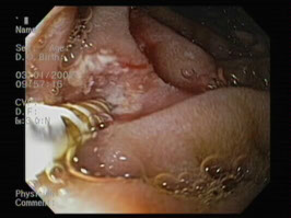
Coagulation of bleeding site
This is a photograph of an area that had been bleeding. The area that had been bleeding is to the left of the center of the photograph. In fact, it had been bleeding quite severely. We used the device, which applies current to the area, to coagulate it, and we can see that there is virtually no bleeding at this point in time. The white that we see in the photograph is where the tissues have been coagulated or burnt, to stop the bleeding. This is one of the main ways we get bleeding sites to stop. You can also see that at the end of the probe, or the straight gold object that is coming out of the endoscope, there appears to be a needle. Once this area was coagulated using heat, which was applied to this probe, I extended a needle out and pierced the tissue with this. I then injected Adrenaline to further halt any bleeding that might occur.
